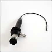
Endoscopy system:
Stryker 988 Endoscope Camera
Storz Rigid Endoscope, Germany
MSI Flexible Endoscope, Germany
Diamond Pro Image Capture Device
Endoscopy permitts the Ear Nose and Throat (ENT) Specialist to see far beyond what is visible with the plain eye. The ENT doctor can navigate through the paranasal sinuses labyrinth, can examine the nasopharynx (far back of the nasal cavity), can visualize the epiglottis, the vocal cords, and can observe and study their function. Visuaization of all the anatomic structures is possible using high magnification and excellent detail, which is very important, especially for the early diagnosis of malignant diseases. Moreover, the endoscopic images and videos can be stored electronically and retieved if necessary in order to compare them with the follow up images.
Endoscopy with the rigid or the flexible endoscope is a safe and painless procedure in the hands of the experienced Ear Nose and Throat Specialist and is allways done in cooperation with the patient.
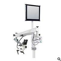
Ear Nose and Throat Microscope
Ecleris Medical - Surgical Microscope
The ear cavity is extremely small in size. With the use of microscope, it is possible to examine the ear cavity in very high magnification. It is extremely useful for the diagnosis of small perforations of the tympanic membrane, retractions of the ear drum, cholesteaotoma, chronic otitis media and otitis media with effusion in children and adults.
Moreover, using the microscope, the trained ENT specialist can perform a variety of in-office procedures, like the correction of a recent tympanic membrane perforation (basic myringoplasty), the insersion of grommets in the ear drum, and the intratympanic injection of medications. Taking sample tissues for biopsies or culture, is facilitated by the use of the microscope.
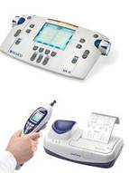
Audiology Equipment
Handheld Tympanometer Easytymp
MA41 Diagnostic Audiometer
from Maico Diagnostic GmbH, Germany.
The diagnosis of hearing loss in adults and children is not an easy task. The measurment of hearing loss, the identification of hearing thresholds using bone conduction and air conduction and the examination of the middle ear function, need extreme detail and the use of nothing but the best of equipment.
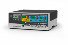
Radiofrequency ablation (RFA)
LED Surtron Flash 160HF Radiofrequency Generator
The Radiofrequency Generator is a highly specilaized instument used for:
LED Surtron Flash 160HF Radiofrequency Generator
The Radiofrequency Generator is a highly specilaized instument used for:
- Inferior nasal turbinate reduction to address chronic nasal obstruction
- "Stiffening" of the soft palate and of the base of tongue to address excessive snoring and mild sleep apnea
- Cryptolysis of the tonsils to address recurrent cryptic tonsillitis assossiated with halitosis (bad smell in mouth)
- Removal of skin lesions (benigh and malignant)
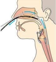
Snoring and Sleep apnea study
The basic cause of snoring and sleep apnea is the obstruction of the airway. This obstuction can happen at any level from the nose to the glottis (the entrance of larynx). The narrowing of the airway causes turbulent flow of the inhaled and exhaled air which is responsible for the characteristic sound of snoring. There are cases that the obstruction of the airway is complete and the breathing during sleep is impossible. If this happens then the individual wakes up in order to breath, which in turn, causes daytime sleepiness and long term problems in the cardiovascular system, through very complex physiologic mechanisms. .
The identification of the exact position of the obstruction of the airway during sleep is of paramount importance in order to offer the correct treatment, surgical or not.
Using careful clinical examination, history taking and endocscopy, identification ot the site of obstuction is possible with a high degree of certainty. Endoscopoy with the flexible endoscope and the review of the recorded images, can give a precise estimation of the site of obstruction.
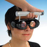
Frenzel gogles
Dehag, Germany
Frenzel gogles are necessary for the clinical and neuro-otologic examination of any patient presenting with dizziness. The removal or optical fixation, magnifies the reflex eye movements that are characteristic of vestibular diseases. The observation of these reflex eye movements enables the Otolaryngologist to precicely locate the site of the disease and to give the appropriate treatment.
